Low dose. Low retention.
Applying Machine Learning to Radiology
Radiologists constantly compare patient images. Whether it is finding small and faintly-enhancing lesions in post-contrast MRI series, or comparing a new MRI study to the patient's prior studies, the task can be inaccurate and time-consuming.
A.I. Analysis, Inc.'s software uses machine learning to assist radiologists in their daily work, automating the tedious aspects of comparing images so radiologists can focus on clinical judgments.
Our Products
eGad™
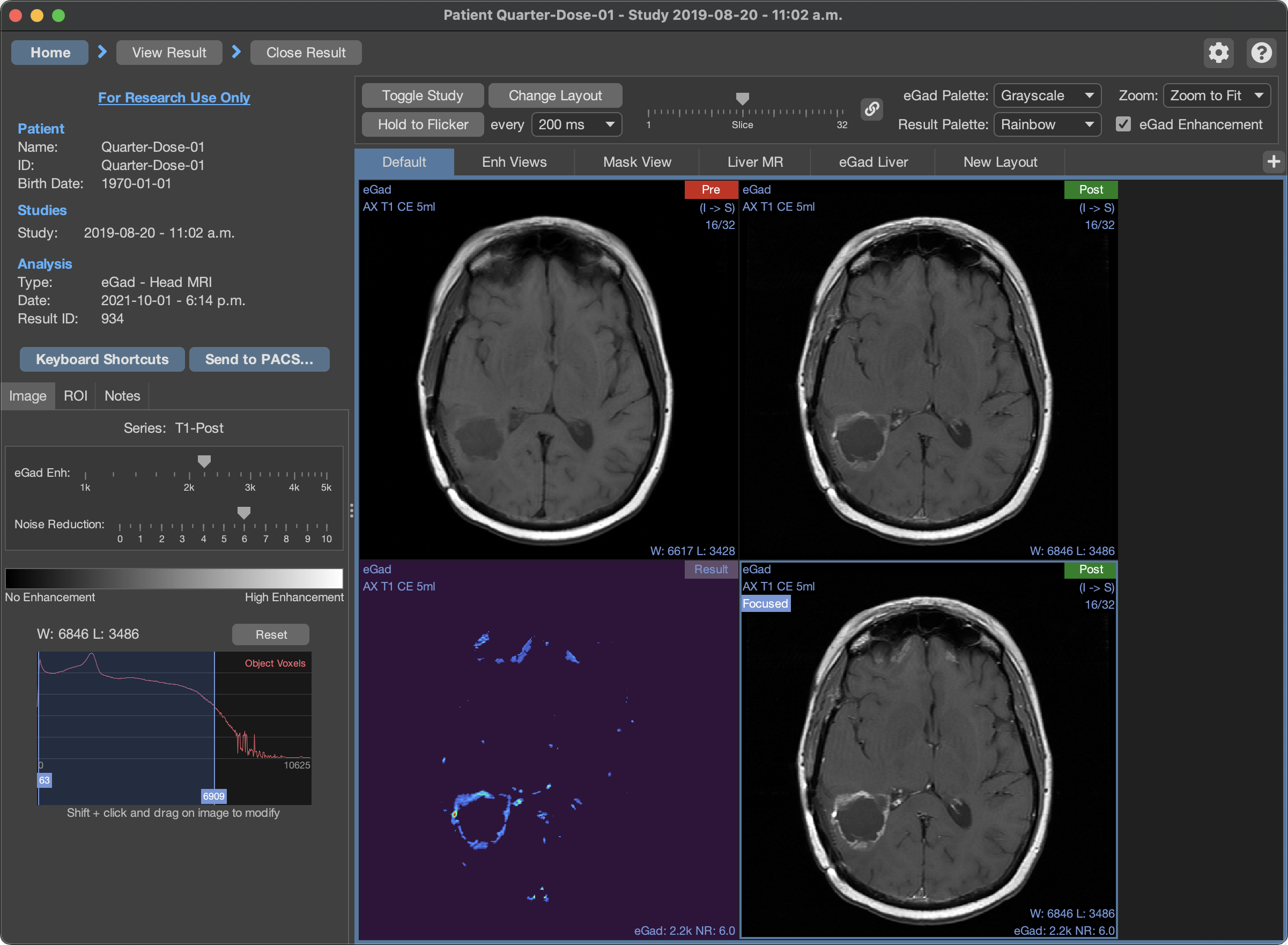
eGad is a suite of tools to make your work with contrast more efficient, more productive, and safer. The eGad machine-learning system intelligently boosts the enhancement in post-contrast MRI images to make enhancing regions more conspicuous to readers.
Learn MoreChange Detector
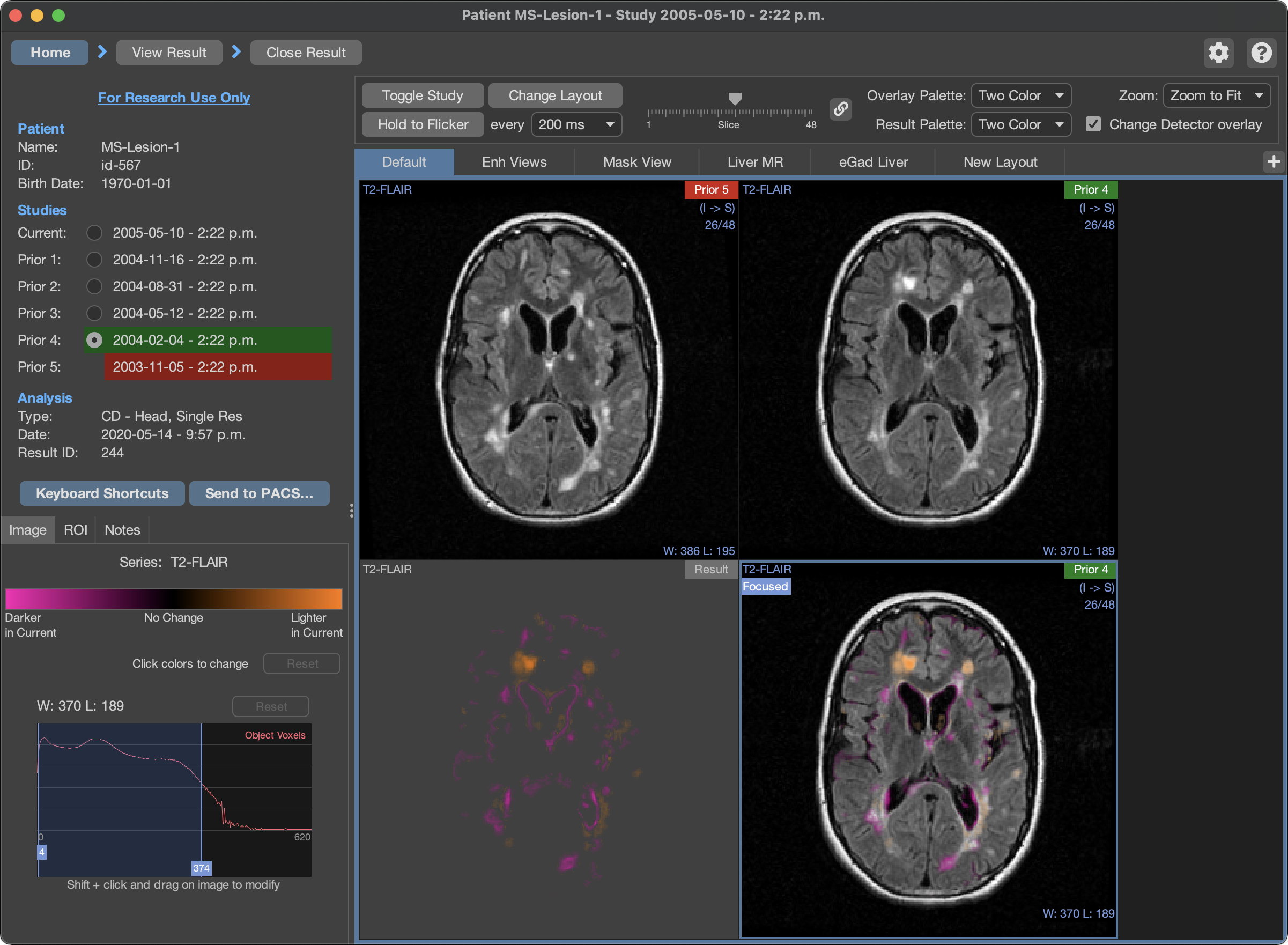
Change Detector is a machine-learning system that compares serial imaging studies, presenting changes between time points as a color-coded overlay indicating what is changing, where, and by what amount.
Learn More