Change Detector
Changes in serial images are hard to see and interpret. A.I. Analysis makes it easy.
The comparison of serial MRI and CT imaging studies is a common task in clinical radiology. Such clinical judgments are, however, not very reproducible. There are a variety of reasons for this, including the confounding of acquisition related changes with disease related changes, and issues related to information presentation.
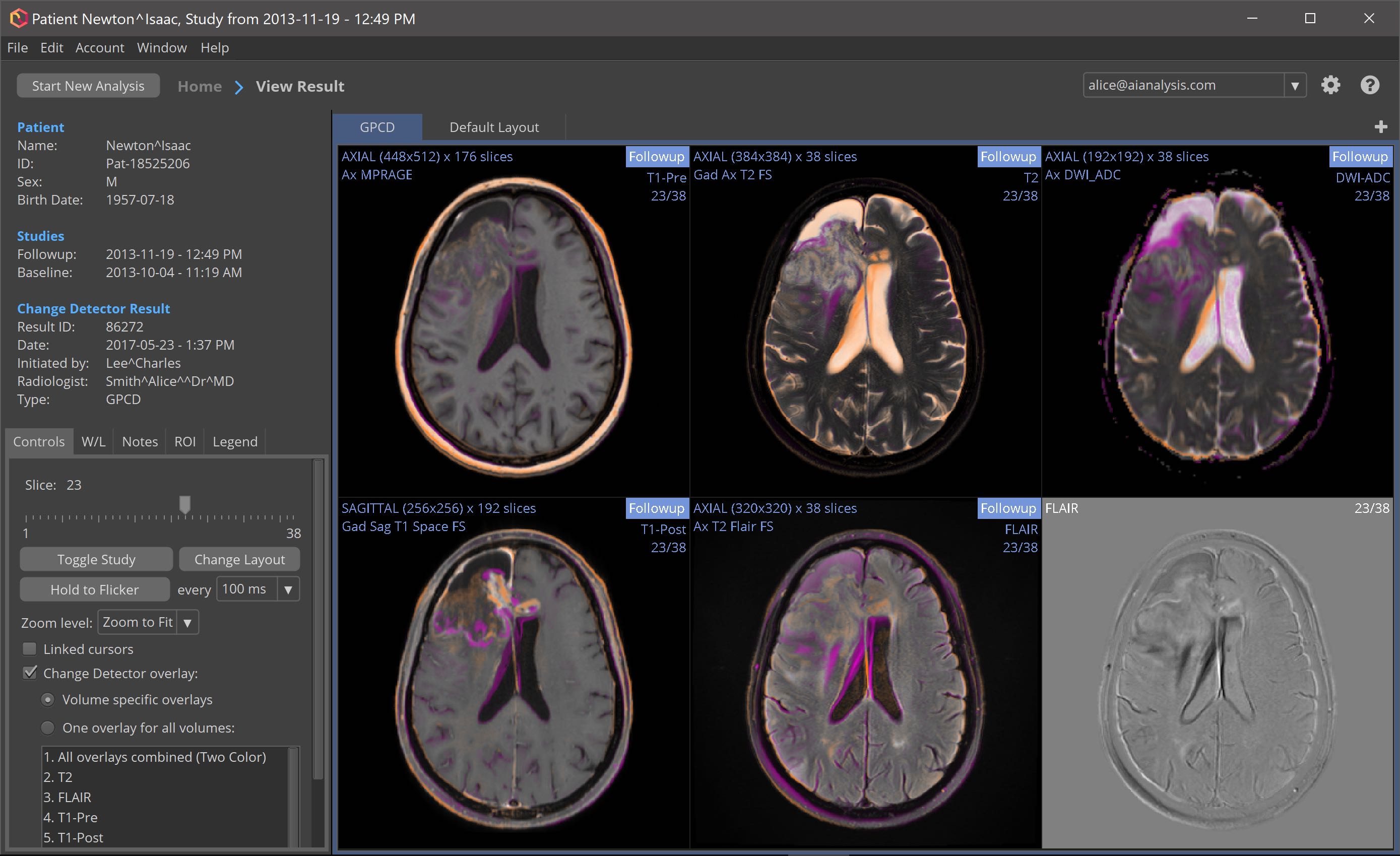
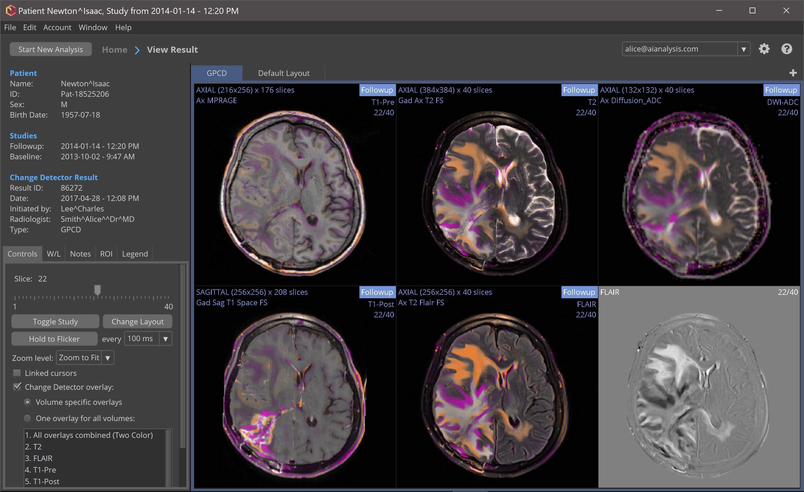
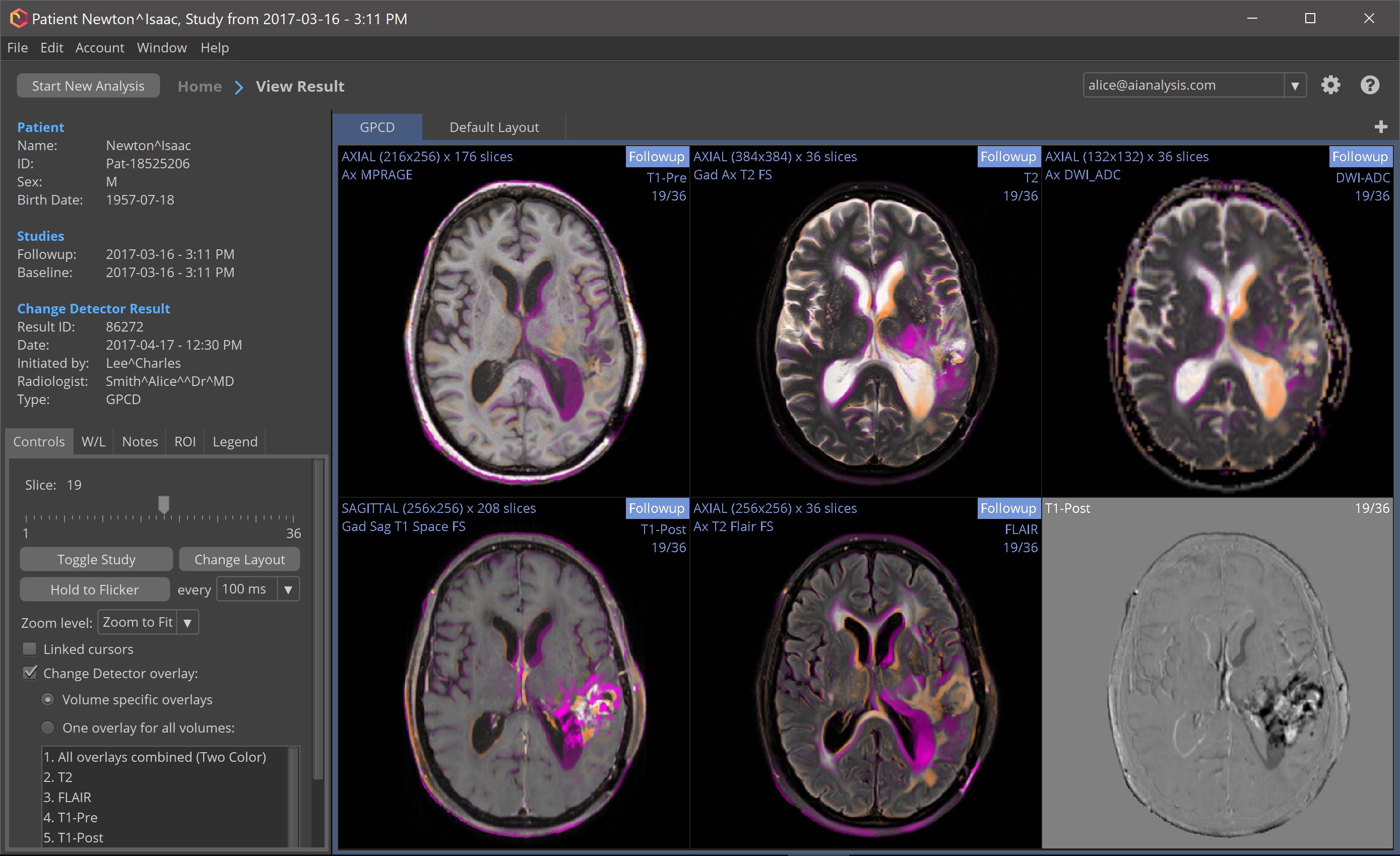
Change Detector is a machine-learning system that compares serial imaging studies, presenting changes between time points as a color-coded overlay indicating what is changing, where, and by what amount. Change Detector reduces information overload and enhances radiologist productivity, increasing value.
Change Detector analyzes the head MRI and CT image types that radiologists use, including T1 (pre- and post-contrast), T2, FLAIR, PD, MTS, ADC, CBV, CBF, MTT, and TTP. Change Detector automatically adapts to new image types, even ones that haven’t been invented yet.
Change Detector demonstrates how practical A.I. systems can free radiologists from the routine task of searching images for changes, making characterization easier, and allowing radiologists to focus on reaching critical judgments and directing therapy.
Automated Change Detection
Change Detector is a software system that compares two or more serial imaging studies, spatially registering prior volumes against the corresponding current volumes of the same type, and presenting changes in the form of a color-coded change map, superimposed on the anatomical images.
Using Change Detector, it may be possible to identify changes months earlier than is possible using manual inspection alone.
Change Detector detects changes in the image types that radiologists use: T1-pre, T1-post, T2, T1-FLAIR, T2-FLAIR, proton density (PD), magnetization transfer suppression (MTS), apparent diffusion coefficient (ADC), cerebral blood volume (CBV), cerebral blood flow (CBF), mean transit time (MTT), and time to peak (TTP). Change Detector is not limited to currently used MRI pulse sequences; it will dynamically adapt to novel image types.
Change Detector is an example of a layered artificial intelligence (AI) system. It demonstrates how practical AI systems can free expert radiologists from routine tasks such as searching images for changing regions, to allow them to focus on reaching critical clinical judgments. The system demonstrates how AI can simply turn information overload into information affluence.
Use Cases
- In the Clinic or Hospital
- Monitoring responses to therapy, and identifying and characterizing disease recurrence. Changes may be found months earlier than they would be using manual inspection alone.
- In Research
- Comparing the efficacy of interventions. Change Detector facilitates large multi-site studies.
- In All Settings
- Results are automatic, quantitative, and reproducible. Given the same input studies, Change Detector will always generate the same analysis results.
Change Detector Strengths
- Change Detector does not require any specific pulse sequences or acquisition parameters. Change Detector works with the images you already acquired. Images being compared can even be different.
- Change Detector can compare images acquired with different scanners.
- Change Detector works with a broad range of diseases.
- Change Detector is quantitative, and can detect and characterize:
- Minimally detectable changes in character
- Minimally detectable changes in shape
The Change Detector desktop application workflow
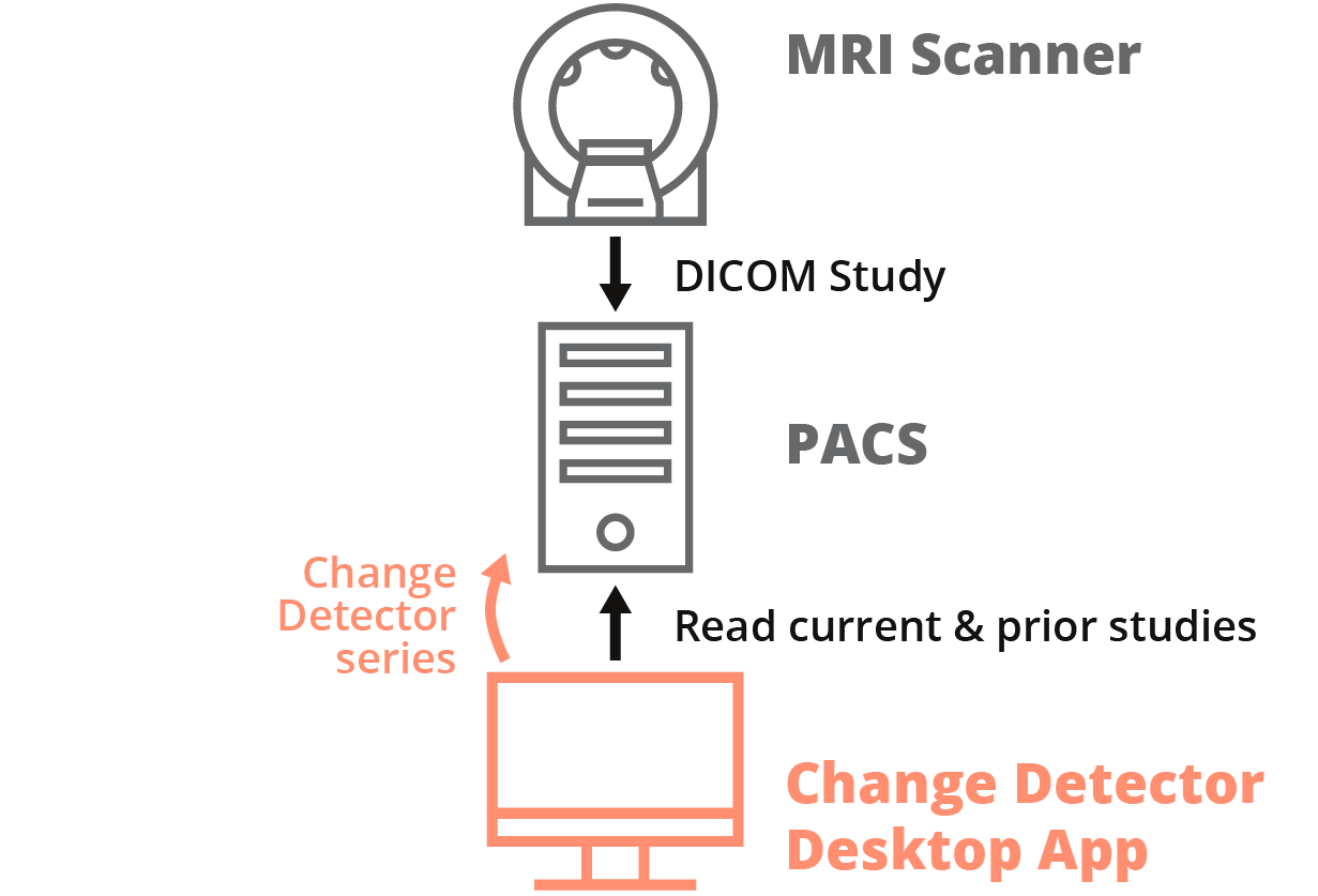
Change Detector post-processing can be performed by a desktop application running on a Windows PC or Mac. A modern laptop or desktop computer with at least 16 GB of RAM is capable of running Change Detector analyses with good performance. Alternatively, Change Detector analyses can be run on a cloud server, providing easy scalability with full security.
- The radiologist retrieves an MRI or CT study from a PACS or from disk, plus one or more prior studies to compare against, and starts the Change Detector analysis.
- The desktop application automatically classifies the different series in the input studies, performs MRI bias field correction, co-registration, Change Detector analysis, and generates Change Detector output series for each input series type.
- The radiologist views the Change Detector result in the desktop application, and optionally sends Change Detector series to a PACS for viewing in the primary viewer.
System Requirements
- Windows 7 or later, or macOS 11 or later
- Intel Core i5 CPU or better (Core i7 CPU recommended)
- At least 8 GB of RAM (16 GB recommended)