eGad™
Small or indistinct regions of contrast enhancement can be hard to detect. A.I. Analysis makes it possible.
eGad is a machine-learning system that intelligently boosts the enhancement in post-contrast MRI images to make enhancing regions more conspicuous to readers.
eGad analysis is available for head MRI and breast MRI, with other areas of anatomy currently being explored. An eGad analysis typically takes 2 to 5 minutes to complete (and can be faster, depending on compute hardware).
Benefits
- A suite of tools to make your work with contrast faster, more efficient, more productive, safer, and more cost-effective
- A Quantitative Enhancement Map (QEM) that shows you what is enhancing, where, and by what amount
- Synthetic Enhancement Boost (SEB): Makes T1-Post enhancing regions more conspicuous, thus enabling greater safety, greater effectiveness, and lower cost
- Objective quantitative measurements, which can be compared over time
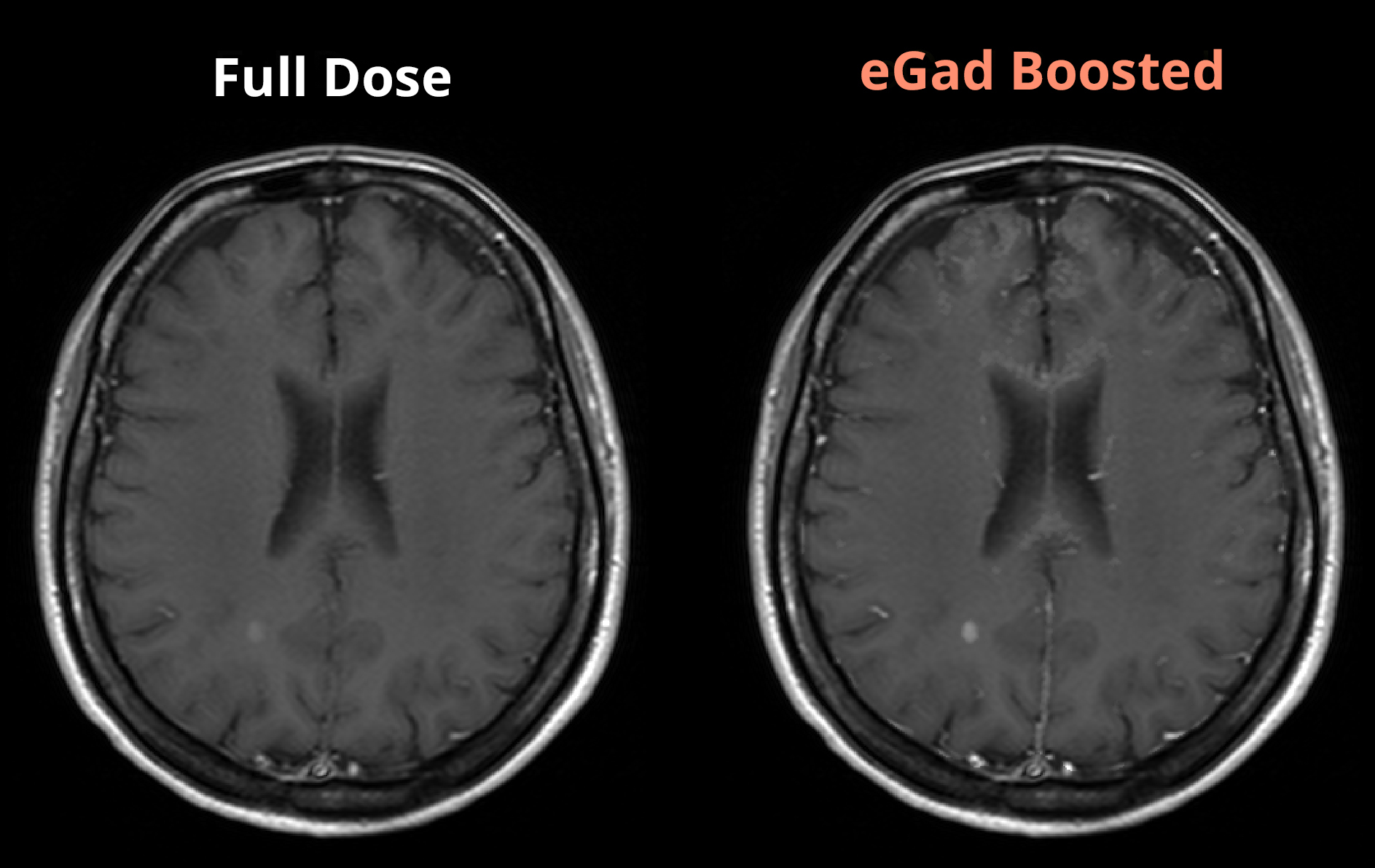
eGad with full dose contrast
eGad can be used to make small and faintly enhancing features more conspicuous. Metastases and multiple sclerosis plaques are easier to see with eGad.
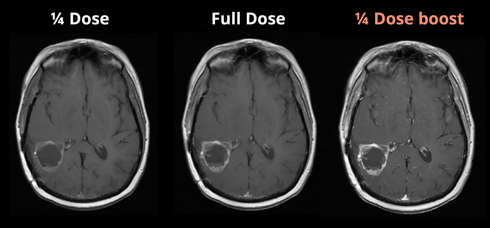
eGad with reduced dose contrast
eGad may also be used in conjunction with a reduced dose of contrast where there are concerns about renal insufficiency or gadolinium retention, such as patients with multiple sclerosis who may be imaged over many years. While a lower contrast dose may produce non-diagnostic image quality, this is remedied by the eGad post-processing, which can produce images with even greater enhancement than a full contrast dose would have provided.
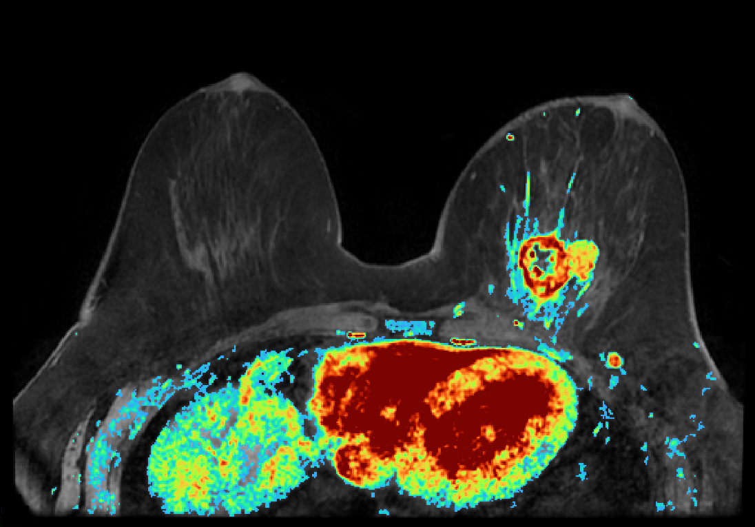
eGad for breast MRI
eGad for breast MRI is a pilot product that measures the kinetic enhancement curve in each voxel of a sequence of post-contrast breast MRI volumes, boosts the conspicuity of enhancing regions, and produces a CAD overlay that highlights abnormalities.
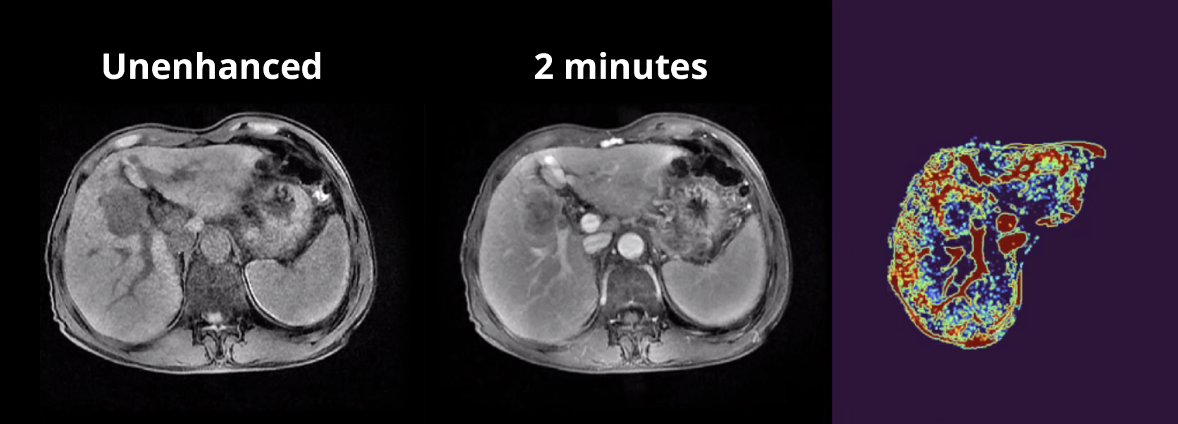
eGad for liver MRI
eGad for liver MRI is a pilot product that measures the kinetic enhancement curve in each voxel of a sequence of post-contrast liver MRI volumes over 10 minutes, and then creates a synthetic 2-hour post-contrast series. This saves time and discomfort for the patient, who does not need to return for a second imaging study two hours after the first, and it frees up time for the MRI suite.
eGad Strengths
- Varying pulse sequences and acquisition parameters, even when there are differences between the Pre- and Post-contrast images
- Varying resolution and orientation, including between the pre- and post-contrast images
- A broad range of contrast agents and doses
- A broad range of disease processes
- A broad range of scanners and practices
eGad uses Genetic Algorithms — a type of artificial intelligence. Like natural evolution, eGad adapts to changing circumstances.
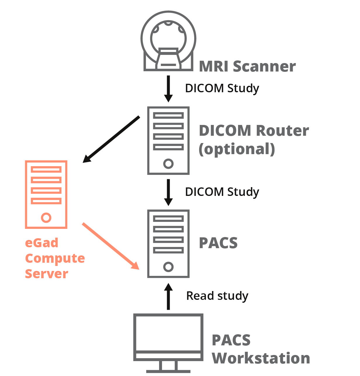
The eGad Compute Server workflow
eGad post-processing can be performed by the eGad Compute Server software running in the hospital or imaging clinic. A general-purpose server running Linux, Windows, or MacOS with at least 16 GB of RAM is capable of running eGad analyses with good performance. Alternatively, eGad analyses can be run on a cloud server, providing easy scalability with full security.
- An MRI scanner completes an MRI study with contrast, and sends the study’s images to the eGad Compute Server directly, or via a DICOM router.
- When the eGad Compute Server receives the MRI study, it automatically classifies the pre- and post-contrast series, performs the eGad analysis, and generates new eGad output series for input post-contrast series.
- The eGad Compute Server sends its output eGad series to a PACS where it can be viewed by the radiologist on any PACS workstation.
System Requirements
- Docker running on Linux, Windows, or macOS
- Intel Core i5 CPU or better (Core i7 CPU recommended)
- At least 8 GB of RAM (16 GB recommended)
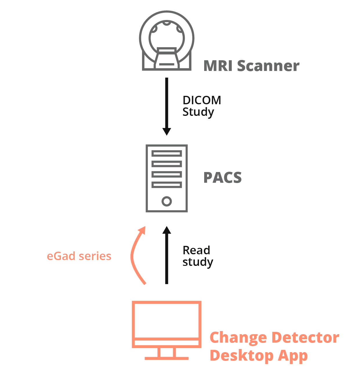
The eGad desktop application workflow
eGad post-processing can be performed by a desktop application running on a Windows PC or Mac. A modern laptop or desktop computer with at least 16 GB of RAM is capable of running eGad analyses with good performance.
- The radiologist retrieves an MRI study with contrast from a PACS or from disk, and starts the eGad analysis.
- The desktop application automatically classifies the pre- and post-contrast series, performs the eGad analysis, and generates new eGad output series for input post-contrast series.
- The radiologist views the output eGad series in the desktop application, and optionally sends eGad series to a PACS for viewing in the primary viewer.
System Requirements
- Windows 7 or later, or macOS 11 or later
- Intel Core i5 CPU or better (Core i7 CPU recommended)
- At least 8 GB of RAM (16 GB recommended)